Nature’s Blueprint: A New Era of Biomedical Innovation from Natural SourcesBy: Suriani, S.Si.,M.Si
Prologue: The Laboratory as a Garden
Part I: The Sea as Scaffold — Marine Collagen and Bacterial Cellulose in Bioprinting
Collagen is the most abundant protein in the human body. It has been determined that this process constitutes the structural framework upon which the integumentary system, the musculoskeletal system, the nervous system, and the endocrine system are organized. For many years, the biomedical industry has relied on collagen extracted from bovine or porcine sources. However, there are potential risks associated with this, including immune reactions, disease transmission and ethical concerns [1].
Concerns have been raised. Marine collagen, derived from sources such as fish skin, scales, and jellyfish, presents a compelling alternative. It shares the same fundamental triple-helix protein structure as mammalian collagen but presents a cleaner immunogenic profile and a more sustainable sourcing pathway. The fish processing industries, which annually discard millions of tonnes of skin and scales, have the potential to become a valuable resource rather than a waste problem [2,3].
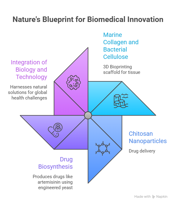
When combined with bacterial cellulose, a nanofibrous material produced by certain bacterial species such as Komagataeibacter xylinus, marine collagen yields a bioink of remarkable quality. Bacterial cellulose is distinguished from plant cellulose in that it is produced as a pure, ultrafine network of fibres, resulting in exceptional tensile strength and water-retention capacity [4,5]. This network functions as the physical backbone of the printed scaffold, while marine collagen provides the biochemical environment that cells recognise as home.
In a 3D bioprinter, the composite material is loaded into a cartridge alongside living cells, then extruded in precise, layer-by-layer formations to construct tissue geometry. The result of the experiment is a scaffold that has been engineered to mimic the extracellular matrix, i.e. the biological environment in which cells are found [6].
Part II: From Shell to Salvation — Chitosan Nanoparticles and Targeted Drug Delivery
Chitosan is notable for its biological properties. The substance under discussion has been found to be biocompatible, a designation that signifies the absence of any adverse bodily reaction to it. It is biodegradable, and upon decomposition, it releases amino sugars that are non-toxic to humans. The substance has been demonstrated to possess antimicrobial properties. It is crucial to note that the substance carries a positive electrical charge at physiological pH. This property gives it an extraordinary affinity for negatively charged cell and mucous membranes, enabling it to adhere, penetrate and release its contents with precision [11].
These properties render chitosan an almost ideal material for use in drug delivery nanoparticles. Nanoparticles are structures measured in nanometers – billionths of a meter – and thus small enough to travel through the bloodstream, navigate biological barriers, and interact with individual cells. The formation of chitosan nanoparticles that encapsulate a drug payload instigates a series of reactions. The nanoparticle's primary function is to protect the drug from degradation in the harsh environment of the bloodstream. The substance under discussion circulates through the body. If engineered with appropriate surface ligands — molecular tags that recognize specific proteins on target cells — it can bind selectively to cancer cells, infected tissues, or inflamed sites, releasing its payload precisely where it is needed [12].
The merits of this approach become evident when juxtaposed with conventional chemotherapy. Conventional cytotoxic pharmaceuticals are administered systemically, thus travelling through the entire body and indiscriminately attacking not only tumour cells, but also healthy tissue. The adverse effects of chemotherapy, including hair loss, nausea and immunosuppression, are a consequence of these drugs' inability to differentiate between cancerous and healthy cells. In contrast, the functionalization of Chitosan nanoparticles with antibodies or peptides that recognize antigens overexpressed on tumour surfaces enables the delivery of lethal concentrations of drugs directly to malignant tissue while sparing healthy cells [13].
Research published in the journal Carbohydrate Polymers has demonstrated the successful delivery of doxorubicin, a potent chemotherapy agent, to breast cancer cells by chitosan nanoparticles. This delivery method has been shown to result in significantly higher tumour accumulation and reduced systemic toxicity in comparison with conventional free drug administration [14]. Furthermore, studies have explored the use of chitosan systems for gene therapy, vaccine delivery, and the treatment of inflammatory bowel disease, where the mucoadhesive properties of chitosan allow prolonged contact with the intestinal mucosa [15].
Part III: The Engineered Yeast — Artemisinin and the Biosynthesis Revolution
For many years, the production of artemisinin was dependent on the cultivation of Artemisia annua, a process which was vulnerable to agricultural unpredictability, climate variation, and the economics of smallholder farming in the developing world. A drought in one season could cause prices of artemisinin to increase, thus rendering life-saving medication inaccessible to populations most in need. The central question guiding this research was whether it was possible to transplant the molecular machinery of Artemisia annua into a more controllable organism [18].
The molecular structure of artemisinin is a remarkable example of chemical complexity, featuring a sesquiterpene lactone with an unusual endoperoxide bridge, which is responsible for its antimalarial activity. This bridge reacts with iron within malaria parasites, generating toxic free radicals that destroy the pathogen from within. Artemisinin can be visualised as a holographic molecular model, with its structure being constituted by a three-dimensional sculpture of carbon rings and oxygen atoms. This intricacy is both beautiful and devastating in terms of its biological effect [19].
Epilogue: Nature and Technology as One
It is estimated that the ocean has invested a considerable amount of time, spanning 500 million years, in the refinement of structural proteins. Crustaceans have evolved exoskeletons that exhibit extraordinary biocompatibility. It is evident that plants have developed chemical arsenals against parasites and pathogens over the course of evolution. In each instance, the solution to the problem at hand was provided by nature itself. The role of modern biomedicine is to understand that solution, deconstruct it to its molecular logic, and rebuild it — in a bioprinter, a nanoparticle, or a fermentation vessel — in service of human health.
This endeavor is not merely an intellectual undertaking; it is a commitment to the pursuit of knowledge and understanding. The magnitude of these stakes is evident in the lives lost: in patients awaiting organs that are not available; in cancer patients subjected to toxic therapies that lack the capacity to differentiate between friend and foe; and in children succumbing to malaria in sub-Saharan Africa due to a lack of access to medication. The convergence of marine biology, polymer chemistry, synthetic biology, and nanotechnology is not merely theoretical; it is the most practical and urgent endeavor of our time.
Reference
1. Browne, S.; Zeugolis, D. I. Collagen?: Finding a Solution for the Source. TISSUE Eng. Part A 2013, 19, 36–39. https://doi.org/10.1089/ten.tea.2012.0721.
2. Coppola, D.; Oliviero, M.; Vitale, G. A.; Lauritano, C.; Ambra, I. D.; Iannace, S.; Pascale, D. De. Marine Collagen from Alternative and Sustainable Sources?: Extraction , Processing and Applications. Mar. Drugs 2020, 18 (214), 1–23.
3. Ahmed, R.; Haq, M.; Chun, B. Characterization of Marine Derived Collagen Extracted from the By-Products of Bigeye Tuna (Thunnus Obesus). Int. J. Biol. Macromol. 2019. https://doi.org/10.1016/j.ijbiomac.2019.05.213.
4. Swingler, S.; Gupta, A.; Gibson, H.; Kowalczuk, M.; Heaselgrave, W.; Radecka, I. Recent Advances and Applications of Bacterial Cellulose in Biomedicine. Polymers (Basel). 2021, 412 (13), 1–29.
5. Utoiu, E.; Manoiu, V. S.; Oprita, E. I.; Craciunescu, O. Bacterial Cellulose?: A Sustainable Source for Hydrogels and 3D-Printed Scaffolds for Tissue Engineering. Gels 2024, 387 (10), 1–20.
6. Nagaraja, K.; Dhokare, P.; Bhattachariyya, A.; Noh, I. Recent Advances in 3D Bioprinting of Polysaccharide-Based Bioinks for Fabrication of Bioengineered Tissues. Mol. Syst. Des. Eng. 2024, 9 (10), 977–999. https://doi.org/10.1039/d4me00001c.
7. Murphy, S. V; Atala, A. Review 3D Bioprinting of Tissues and Organs. Nat. Publ. Gr. 2014, 32 (8), 773–785. https://doi.org/10.1038/nbt.2958.
8. Vijayavenkataraman, S.; Yan, W.; Feng, W.; Wang, C.; Ying, J.; Fuh, H. 3D Bioprinting of Tissues and Organs for Regenerative Medicine ?. Adv. Drug Deliv. Rev. 2018, 132, 296–332. https://doi.org/10.1016/j.addr.2018.07.004.
9. Silva, T. H.; Moreira-Silva, J.; Marques, A. .; Domingues, A.; Bayon, Y.; Reis, R. L. Marine Origin Collagens and Its Potential Applications. Mar. Drugs 2014, 5881–5901. https://doi.org/10.3390/md12125881.
10. Kou, S.; Peters, L.; Mucalo, M. Chitosan: A Review of Sources and Preparation Methods. Int. J. Biol. Macromol. 2020. https://doi.org/10.1016/j.ijbiomac.2020.12.005.
11. Younes, I.; Rinaudo, M. Chitin and Chitosan Preparation from Marine Sources. Structure, Properties and Applications. Mar. Drugs 2015, 1133–1174. https://doi.org/10.3390/md13031133.
12. Masood, F. Polymeric Nanoparticles for Targeted Drug Delivery System for Cancer Therapy. Mater. Sci. Eng. C 2016, 60, 569–578. https://doi.org/10.1016/j.msec.2015.11.067.
13. Mitragotri, S.; Burke, P. A.; Langer, R. Overcoming the Challenges in Administering Biopharmaceuticals: Formulation and Delivery Strategies Samir. Nat. Rev. Drug Discov. 2015, 13 (9), 655–672. https://doi.org/10.1038/nrd4363.Overcoming.
14. Bhattarai, N.; Gunn, J.; Zhang, M. Chitosan-Based Hydrogels for Controlled , Localized Drug Delivery ?. Adv. Drug Deliv. Rev. 2010, 62 (1), 83–99. https://doi.org/10.1016/j.addr.2009.07.019.
15. Antoniou, V.; Mourelatou, E. A.; Galatou, E.; Avgoustakis, K.; Hatziantoniou, S. Gene Therapy with Chitosan Nanoparticles?: Modern Formulation Strategies for Enhancing Cancer Cell Transfection. Pharmaceutics 2024, 1–28.
16. Li, J.; Cai, C.; Li, J.; Li, J.; Li, J.; Sun, T.; Wang, L.; Wu, H.; Yu, G. Chitosan-Based Nanomaterials for Drug Delivery. Molecules 2018, 1–26. https://doi.org/10.3390/molecules23102661.
17. White, N. . Qinghaosu (Artemisinin): The Price of Success. Science (80-. ). 2008, 330. https://doi.org/10.1126/science.1155165.
18. Paddon, C. J.; Keasling, J. D. Gy Semi-Synthetic Artemisinin?: A Model for the Use of Synthetic Biology in Pharmaceutical Development. Nat. Rev. 2014, 12 (355–367). https://doi.org/10.1038/nrmicro3240.
19. Meshnick, S. R. Artemisinin?: Mechanisms of Action , Resistance and Toxicity. Int. J. Parasitol. 2002, 32, 1655–1660.
20. Galanie, S.; Thodey, K.; Trenchard, I. j; Interrante, M. F.; Smolke, C. D. Complete Biosynthesis of Opioids in Yeast. Science (80-. ). 2016, 349 (6252), 1095–1100. https://doi.org/10.1126/science.aac9373.Complete.